A 2D Echocardiogram with Color Doppler is an advanced, non-invasive diagnostic technique used to assess the structure and function of the heart. This imaging method combines traditional 2D ultrasound with Color Doppler technology, providing real-time, high-resolution images of the heart’s chambers, valves, and blood vessels, while simultaneously visualizing blood flow.
How It Works:
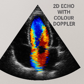
2D Echocardiogram: This part of the test creates two-dimensional cross-sectional images of the heart. These images provide clear visualizations of the heart's anatomy, including the walls, valves, and major blood vessels. The 2D images help doctors assess the size, shape, and function of the heart.
Color Doppler: The Color Doppler feature adds a dynamic dimension by color-coding the movement of blood within the heart. Red and blue hues are used to indicate the direction of blood flow—red shows blood flowing towards the transducer (probe), while blue represents blood moving away. This feature is crucial for identifying abnormal blood flow patterns, such as those caused by valve issues, heart defects, or blockages in the arteries.
Clinical Applications:
Valvular Heart Disease: Color Doppler helps detect abnormalities in heart valves, such as stenosis or regurgitation, by visualizing blood flow through the valves.
Congenital Heart Defects: It aids in identifying structural heart abnormalities present from birth.
Cardiac Function: It evaluates the efficiency of the heart's pumping action and can be used to assess conditions such as heart failure.
Detecting Blood Clots or Blockages: Color Doppler can identify abnormal blood flow patterns caused by blockages, thrombosis, or narrowing of the blood vessels.
Benefits:
Non-invasive: No incisions or injections are required, making it a safe and pain-free procedure.
Quick Results: The procedure is typically fast, and results are available almost immediately.
High Accuracy: Color Doppler provides highly accurate and clear images, enabling doctors to make informed decisions about treatment options.
Real-time Imaging: It allows for immediate visualization of blood flow and heart function during the procedure.
A 2D echocardiogram with color Doppler is a powerful diagnostic tool that provides vital insights into the health of the heart, allowing for early detection and management of various cardiac conditions. It is commonly used in both routine heart checkups and as part of the diagnostic process for patients experiencing symptoms of heart disease.
 Go to homepage